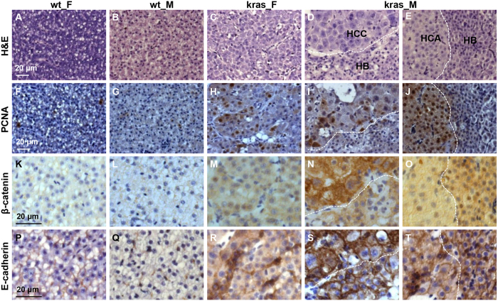
Figure 4. Characterization of the multi-nodular liver tumor from male krasV12 zebrafish after long-term tumor induction.
Adjacent liver sections in female and male wild type and krasV12 fish at 5 mpi were processed for H&E and immunohistological staining. The multi-nodular male tumors have mixed types of tumors (HCC, HB and HCA) which were separated by dash lines. (A–E) H&E staining. (F–J) Immunocytochemical staining for PCNA. (K–O) Immunohistochemical staining for β-catenin. (P–T) Immunohistochemical staining for E-cadherin. Scale bar, 20 μm for all panels.

