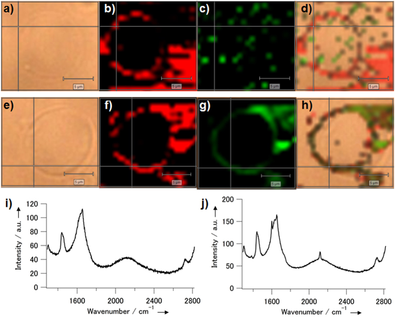
Figure 3. Raman microscopic imaging of the liposomes including the chemically-activatable alkynyl steroid analogue probe before and after activation.
Microscopic images of the liposomes including 1 were obtained (a–d) before and (e–h) after incubation with TsNHNH2. The light field images (a and e), the Raman scattering images at 1400 cm−1 (b and f) and at 2120 cm−1 (c and g), and their merged images (d and h) are shown. Scale bars: 5 μm. (i and j) Raman spectra obtained at the cross point on the liposome membrane in (a) and (e), respectively.

