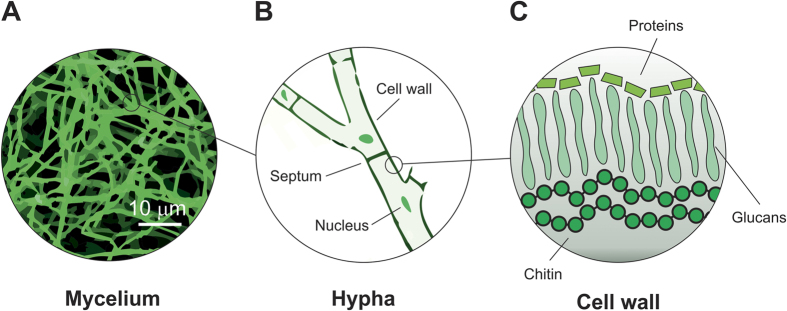
Figure 1. Schematic representation of mycelium physiology at different scales.
(A) Optical microscopy image of a mycelium film showing a branched network of micro-filaments (hyphae). (B) Schematic representation of a hypha that is formed by cells separated by cross walls (septa), all enclosed within a cell wall. (C) Schematic representation of the cell wall that is composed of a layer of chitin on the cell membrane, a layer of glucans (whose composition varies between species) and a layer of proteins on the surface (adapted from ref. 36).

