
Fig. 2.
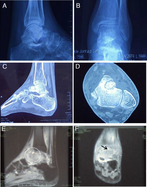
Preoperative imaging of the left ankle prior to the second surgery. (a, b) Preoperative radiographs show pathologic damage to the anterolateral talus, Approximately 1.0-cm-diameter higher-density nodules can be seen on the neck of the talus on the left side, with a clear boundary. (c, d) Preoperative computed tomography images show the characteristics of the osteoid osteoma nidus. (e, f) Preoperative sagittal T2-weighted and coronal T2-weighted magnetic resonance images show widespread bone marrow oedema of the talus with the osteoid osteoma lesion inside (white circle and black arrow indicate the nidus)