Fig. 3.

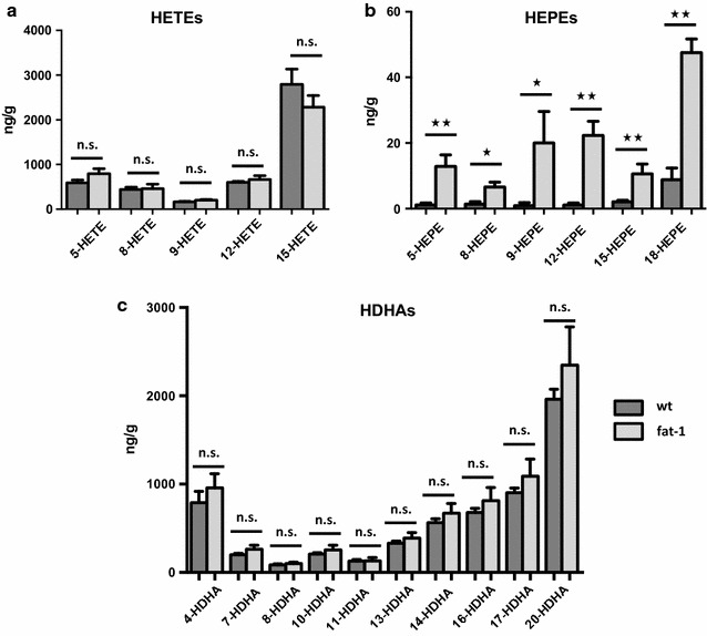
Comparison of lipid metabolite profiles in brain tissue of wt and fat-1 mice. a Levels of AA-derived HETEs were analysed using LC MS/MS. They were relatively similar among wt and fat-1 animals (presented as mean values and SEM). b The only significant difference in lipid metabolites between wt and fat-1 brain tissue was observed for EPA-derived monohydroxy metabolites. Levels of EPA-derived HEPEs were significantly higher in fat-1 animals with particularly high amounts of anti-inflammatory 18-HEPE. c Concerning DHA-derived HDHAs, levels were also relatively similar in the brain tissue of wt and fat-1 mice with no significant differences among any of the DHA-derived metabolites
