Fig. 1.

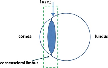
The laser exposed surface of eye from limbus vertically, sequential images with step length of 5 μm were then collected through the trans-scleral imaging. Green dotted box corresponds to the imaging area

The laser exposed surface of eye from limbus vertically, sequential images with step length of 5 μm were then collected through the trans-scleral imaging. Green dotted box corresponds to the imaging area