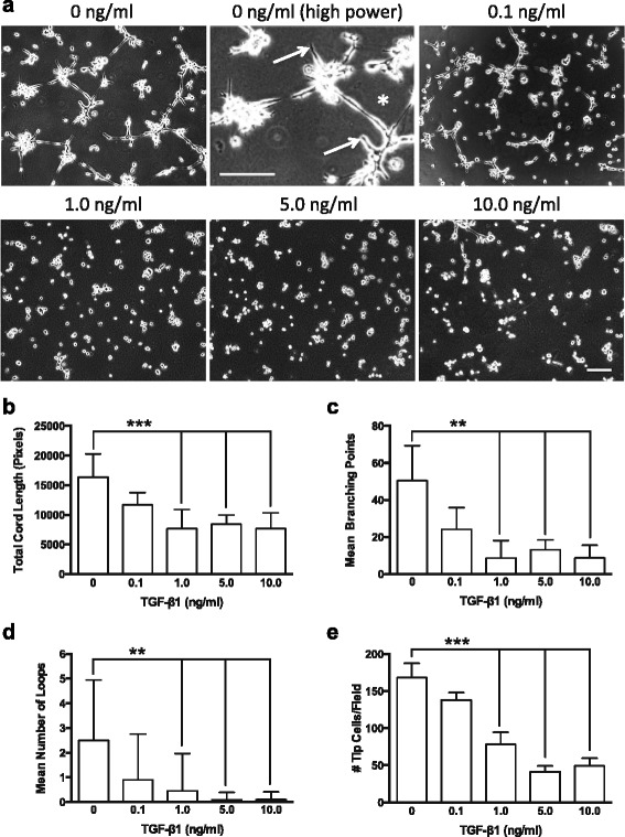
Fig. 1.

TGF-β1 induced dose-dependent reduction in endothelial cord formation. a Representative phase contrast images showing endothelial cell cord formation 8 h after plating on Matrigel™ in the presence of exogenous TGF-β1 (0–10 ng/ml). Note higher magnification control panel (0 ng/ml TGF-β1), showing tip cells (arrows) and loop of endothelial cords (asterisk). WimTube automated quantification of cord formation showed that TGF-β1 significantly inhibits total cord length (b), cord branching (c), formation of loops (d), and generation of tip cells (e). TGF-β1 doses of 1.0 ng/ml or higher significantly inhibited cord formation compared to control (0 ng/ml) or 0.1 ng/ml. **p ≤ 0.01; **p ≤ 0.001; N = 4; Kruskal-Wallis test. Scale bars = 300 μm
