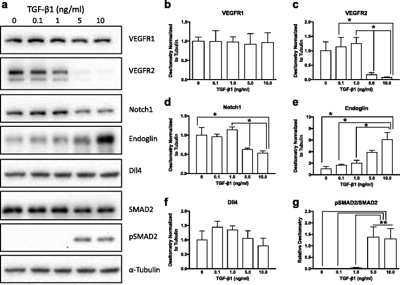
Fig. 2.

TGF- β1 induced dose-dependent changes in endothelial cell receptors. a Representative western blots of endothelial cells exposed to exogenous TGF-β1 (0.1–10.0 ng/ml) for 24 h. Densitometry showed significantly reduced VEGFR2 expression (b), but no change in VEGFR1 (c) in cells treated with higher doses of TGF-β1. The TGF-β1 co-receptor endoglin was significantly upregulated in a dose dependent fashion (e). Notch1 was significantly downregulated in endothelial cells treated with 5.0 and 10.0 ng/ml TGF-β1 (d), and its ligand Dll4 showed a trend towards reduced expression with higher doses (f). TGF-β1 signaling through ALK5/SMAD2-dependent pathways, as revealed by phosphorylation of SMAD2, was maximal at doses of 5.0 ng/ml and higher (g). *p ≤ 0.05; **p ≤ 0.01; N = 3; Kruskal-Wallis test
