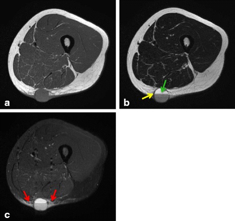
Fig. 3.

A 28-year-old woman diagnosed with angiomatoid fibrous histiocytoma (case 1): (a) T1-weighted spin echo, (b) T2-weighted spin echo, and (c) STIR images. A 21 × 23 × 22-mm well-circumscribed, round mass is present in the subcutaneous fat of the posterior right thigh. The lesion is homogeneously hypointense on T1 WI and presents fluid–fluid level (green arrow) and pseudocapsule (yellow arrow) on T2 WI. STIR shows peritumoral edema (red arrows)
