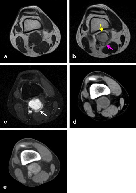
Fig. 4.

A 36-year-old man diagnosed with angiomatoid fibrous histiocytoma (case 5): (a) T1-weighted spin echo, (b) T2-weighted spin echo, and (c) contrast-enhanced MR images. (d) Non-enhanced and (e) enhanced CT images. A 32 × 36 × 45-mm asymptomatic mass is present in the popliteal lesion of right knee. The lesion is homogeneously isointense on T1 WI and presents with a multilocular area (pink arrow) and pseudocapsule (yellow arrow) on T2 WI. A contrast-enhanced MR image shows intratumoral and peritumoral (white arrow) enhancement. In addition, an enhanced CT image shows variegated enhancement
