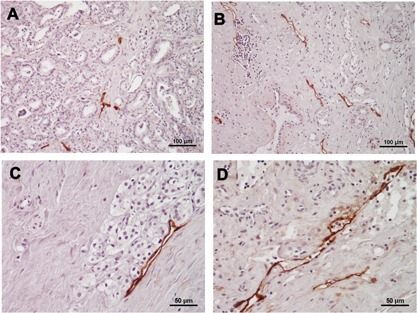
Figure 1.

Representative micrographs of D2‐40‐positive lymph vessels in prostate cancer tissue samples with/without NHT. Most lymph vessels were relapsed and the intra‐luminal space was narrow in non‐NHT specimens (A: magnification ×200). Lymph vessels had a relatively wide inner cavity in NHT specimens (B: magnification ×200, C: ×400). Some cells were detected within D2‐40‐positive lymph vessels in some NHT specimens (D: magnification ×400).
