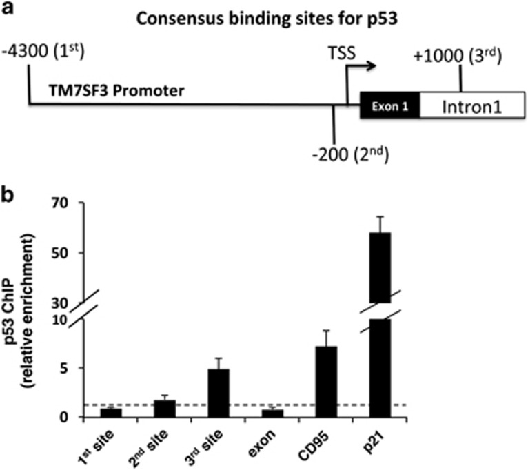
Figure 7.
Chromatin immunoprecipitation (ChIP) of p53 in MCF10A cells. MCF10A cells were subjected to ChIP with p53-specific polyclonal antibodies (CM1) or no antibody control (mock). Quantification of the fragment containing the predicted p53-binding site in the TM7SF3 (site 1 – 4300 bp and site 2 – 180 bp upstream to the TSS; site 3 – 1000 bp downstream (first intron) to the TSS); CD95, or the p21 promoter regions was done by qRT-PCR; dashed line – Mock. Bar graphs are the mean±S.E.M. of three experiments

