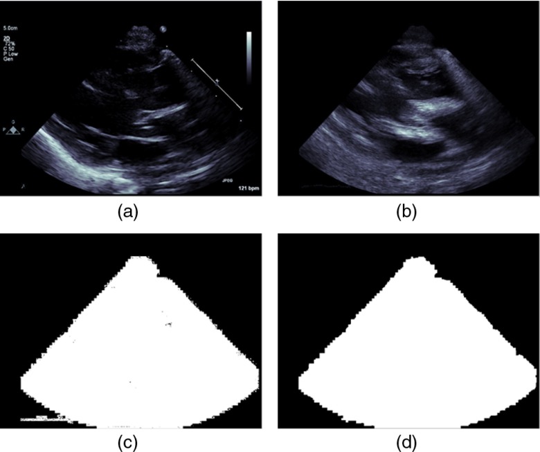
Fig. 7.
Process for obtaining the image acquisition mask from annotated image data: (a) frame of 2-D echocardiographic temporal loop data in the PSLax view (b) the sum of the frame-wise differences are then computed over an even number of frames, (c) the binary mask corresponding to the nonzero locations of the sum image are then determined, (d) pixels corresponding to the maximal component of the connected components algorithm applied to (c) to get the data specific acquisition mask.

