
Figure 1.
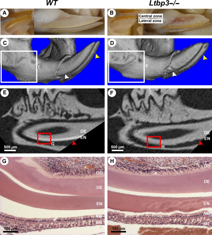
External morphology, micro‐computed tomography (micro‐CT), and histology analysis of lower incisors of a 3.5‐month‐old wild‐type (WT) mouse (left column of panels) or a latent transforming growth factor beta binding protein mutant (Ltbp3‐/‐) mouse (right column of panels). The same WT or Ltbp3‐/‐ mutant is shown in all respective panels. (A, B) In external views of lower incisors, the incisors of the mutant are shorter, laterally white, and centrally orange. (C, D) Three‐dimensional rendered micro‐CT images of the distal part of the mandible. The surface of enamel from the Ltbp3‐/‐ mouse appears irregular (yellow arrows), and enamel nodules are seen near the alveolar bone crest area of the incisor (white arrows). (E, F) Two‐dimensional micro‐CT sagittal sections of the first lower molar and the underlying incisor (obtained from the respective boxed region in panels C and D), showing the enamel density. In the mutant the enamel layer is clearly reduced and has a rough appearance (red arrows). (G, H) Histological sections (hematoxylin and eosin staining) of the lower incisor (respective red boxed area in panels E and F) showing an irregular enamel layer (EN) in the Ltbp3‐/‐ mutant. The odontoblast (OD) and dentin (DE) layers show no obvious changes. AM, ameloblasts; SR, stellate reticulum.