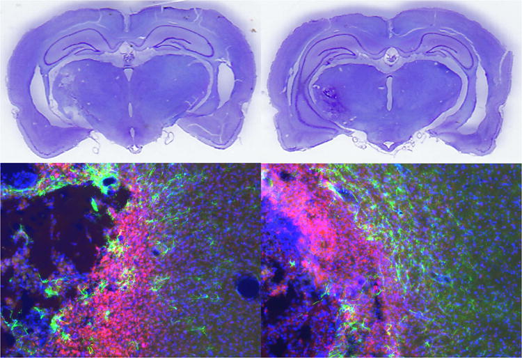
Figure 7.

Representative histological images of a control rat (left) and grafted rat (right, group 4W). The upper images are cresyl violet stained brain sections showing subcortical infarction. The lower images are immunohistochemistry of the infarct showing astrocytosis (GFAP in green), microcytosis (CD11B in red), and overall cellularity (Hoechst in blue).
