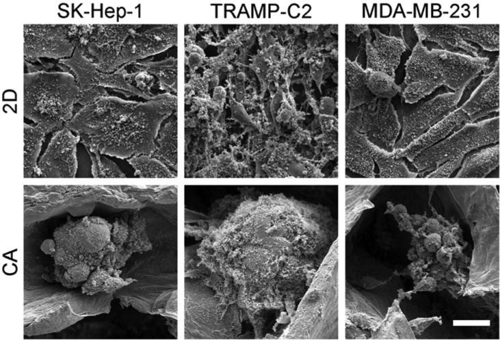
Figure 3.
SEM images of SK-Hep-1 (hepatocellular carcinoma), TRAMP-C2 (prostate cancer), and MDA-MB-231 (breast cancer) cells cultured in 2D well plates and CA scaffolds at day 10. Cells cultured on 2D plates have a more epithelial-like appearance whereas cells cultured in CA scaffolds have a more rounded appearance and form tumor spheroids. Scale bar represents 20 μm.

