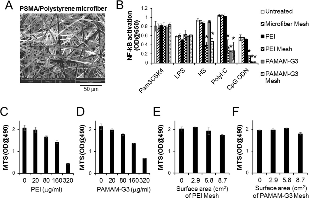
Fig. 1.
NABP-immobilized PSMA/polystyrene microfiber mesh scavenges multiple TLR agonists without cytotoxicity. A. scanning electron microscope (SEM) image of surface of PSMA/polystyrene microfiber mesh. B. Complete culture media (1 ml) supplemented with TLR agonists Pam3CSK4, LPS, Heparan sulfate (HS), polyI:C or CpG ODN, were incubated for 1 min with or without either PEI- or PAMAM-G3-immobilized microfiber mesh (2.9 cm2) at room temperature. The treatment was repeated once. TLR reporter cells were incubated for 3 days in either untreated or treated complete culture media. For the treatment with free NABPs, TLR reporter cells in the complete culture media containing fetal bovine serum and TLR agonist were directly treated with either PEI (20 µg/ml) or PAMAM-G3 (25 µg/ml). The activation of NF-κB in TLR signaling pathway was determined by a colorimetric enzyme assay. C-F. Human primary fibroblasts were cultured in either complete culture media containing free (C) PEI or free (D) PAMAM-G3 at various concentrations or complete culture media pre-treated with (E) PEI-immobilized mesh or (F) PAMAM-G3-immobilized mesh at various surface areas (0.288 mg/cm2 PEI on mesh; 0.128 mg/cm2 PAMAM-G3 on mesh). After 3 days incubation, cell proliferation was determined by a MTS (3-(4,5-dimethylthiazol-2-yl)-5-(3-carboxymethoxyphenyl)-2-(4-sulfophenyl)-2H–tetrazolium) assay. Error bars are S.D.. * Significant different (P < 0.05), vs untreated group.

