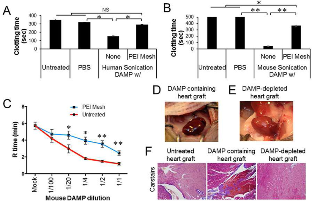
Fig. 5.
Inhibition of DAMP-induced clotting by PEI-immobilized PSMA/polystyrene microfiber mesh. Human and mouse sonication-induced DAMPs were incubated for 1 min with or without PEI-immobilized PSMA/polystyrene microfiber meshes (2.9 cm2). The treatment was repeated four times. A, B. Platelet-depleted plasma from human and mouse normal blood were incubated with the DAMPs (10% v/v) and the clotting time measured. C. Mouse whole blood was incubated with the DAMPs at various dilutions with PBS. The coagulation (R) time was detected by Thromboelastography (TEG). D, E. Donor hearts isolated from normal mice (n=3) was perfused with the DAMP (2 ml), followed by heterotrophic heart transplantation. Heart beating and thrombosis of donor heart was monitored. The image was captured 30 min after unclamping. F. Representative sections from untreated, DAMP-treated, and PEI-immobilized mesh-filtered DAMP-treated allografts harvested 30 min after unclamping. All were stained with Carstairs (modified Masson’s trichrome) to visualize platelets (purple), erythrocytes (clear yellow/orange), and fibrin (Bright red/orange) taken at x100 magnification. Data represent three independent experiments. Error bars are S.D.. NS: statistically non-significant. * P < 0.05. ** P < 0.01.

