Figure 5.
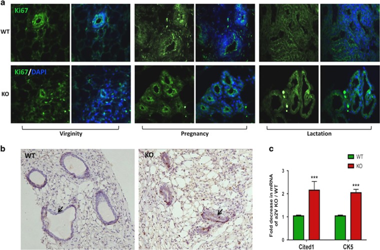
a2V ablation leads to disintegrated mammary epithelium. Inguinal mammary glands from WT and KO mice were evaluated for epithelial lumen integrity and proliferation. (a) Immunofluorescence staining of proliferation marker Ki67 (green) in mammary epithelia of WT and KO mice from virginity, pregnancy and lactation stages. Nucleus is stained by DAPI (diamidino-2-phenylindole; blue). (b) Representative images from immunohistochemistry highlighting constricted lumen (black arrows) in virgin a2V-KO mice processed for incorporation of 5-bromo-2′-deoxyuridine (BrdU). Brown staining – 3,3′ diaminobenzidine (DAB). Original magnification: × 200. (c) Quantitative real-time-PCR (qRT-PCR) shows fold decrease in mRNA expression levels of mammary epithelial cell markers Cited 1 and Keratin 5. Before fold-change calculation, the values were normalized to endogenous control glyceraldehyde 3-phosphate dehydrogenase (GAPDH). Data represent mean±S.E., n=6. ***P≤0.001

