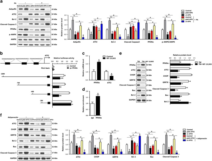
Figure 7.
PPARα inhibited transcription of ATF2 and ER stress-induced apoptosis of adipocytes. (a) Protein levels of adipoR1, ATF2, Bcl-2, cleaved-caspase 3 and PPARα, p-AMPK in adipocytes infected with pAd-APN or si-APN and incubated with PA (n=3). (b) Dual-luciferase reporter assay of ATF2 and PPARα. Cells were transfected with PGL3-basic or PGL3-ATF2 plasmids, and pc-PPARα plasmid (n=3). (c) mRNA levels of PPARα and ATF2 after WY-14 643 treatment (n=3). (d) ChIP analysis between ATF2 and PPARα (n=3). (e) Protein levels of PPARα, ATF2, CHOP, GRP78, cleaved-caspase 3, Bax and Bcl-2 incubated with TM, followed with WY-14 643 (n=3). (f) Protein levels of ATF2, CHOP, GRP78, Bcl-2, Bax and cleaved-caspase 3 of adipocytes infected with pAd-ATF2 or si-ATF2, then incubated with adiponectin (n=3). pAd-APN, recombinant adenovirus overexpression vector of adiponectin; si-APN, recombinant lentiviral interference vector of adiponectin. pAd-ATF2, recombinant adenovirus overexpression vector of ATF2; si-ATF2, recombinant lentiviral interference vector of ATF2. Values are means±S.D. *P<0.05 compared with the control group

