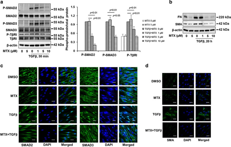
Figure 1.
MTX attenuates TGFβ-1 signaling in human lung fibroblast. (a) MRC5 cells were treated with increasing doses of MTX (0, 1, 5, 10 μM) for 1 h, and then cells were treated with TGFβ-1 (2 ng/ml) for 30 min; the phosphorylated and total forms of SMAD2, SMAD3, and TβRI were then analyzed by western blotting. Western blotting images were cropped to improve the conciseness of the data; samples derived from the same experiment and the blots were processed in parallel. Representative of experiments performed at least three independent times. Intensities of blots were measured. The ratio of phosphorylated/total protein was analyzed by the ImageJ software. (b) MRC5 cells were treated with increasing doses of MTX (0, 1, 5, 10 μM) for 1 h, and then cells were treated with TGFβ-1 (2 ng/ml) for 20 h. FN and SMA levels were analyzed by western blotting. Western blotting images were cropped to improve the conciseness of the data; samples derived from the same experiment and the blots were processed in parallel. Representative of experiments performed at least three independent times. (c) MRC5 cells grown on glass-bottom dishes were treated with dimethyl sulfoxide (DMSO) or MTX (5 μM) for 1 h, and then cells were treated with TGFβ-1 (2 ng/ml) for 1 h. Localization of SMAD2 or SMAD3 (green) in the cells were detected by immunostaining with SMAD2 and SMAD3 antibodies. DAPI (4,6-diamidino-2-phenylindole) was used for nuclei staining (blue). Bars, 10 μM. Representative images were shown. (d) MRC5 cells grown on glass-bottom dishes were treated with DMSO or MTX (5 μM) for 1 h, and then cells were treated with TGFβ-1 (2 ng/ml) for 20 h. Localization of SMA (green) in the cells were detected by immunostaining with an SMA antibody. DAPI was used for nuclei staining (blue). Bars, 10 μm. Representative images are shown

