Figure 4.
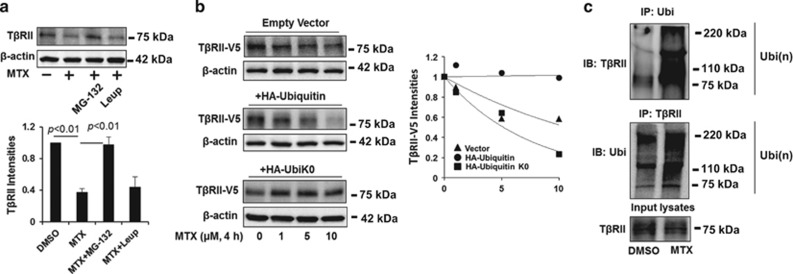
MTX promotes TβRII ubiquitination and degradation in the proteasome. (a) MRC5 cells were treated with MG-132 (20 μM) or leupeptin (100 μM) for 1 h prior to MTX treatment (5 μM, 2 h). TβRII and β-actin levels were analyzed by western blotting. Intensities of TβRII were analyzed by the ImageJ software. (b) MRC5 cells were co-transfected with TβRII-V5 and with either empty vector, HA-ubiquitin, or HA-ubiquitin without lysine (HA-UbiK0) plasmids, cells were then treated with increasing concentrations of MTX (0–10 μM) for 4 h. TβRII-V5 and β-actin levels were analyzed by western blotting. Intensities of TβRII-V5 were analyzed by the ImageJ software and then compared between the three groups. (c) MRC5 cells were treated with 5 μM MTX for 1 h, and then cell lysates were subjected to immunoprecipitation with an ubiquitin antibody or a TβRII antibody, followed by TβRII or ubiquitin immunoblotting. Input lysates were analyzed by TβRII immunoblotting. Western blotting images were cropped to improve the conciseness of the data; samples derived from the same experiment and the blots were processed in parallel. Representative of experiments performed at least three independent times

