Figure 1.
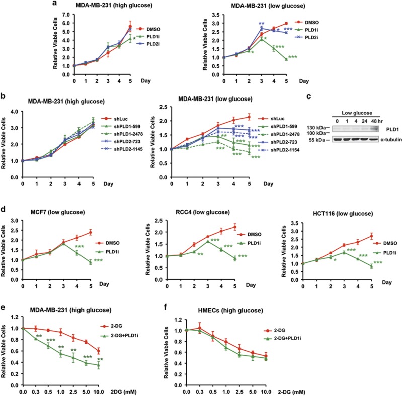
Requirement of PLD activity for cancer cell survival in low glucose medium. (a) PLD1 and PLD2 small-molecule inhibitors, PLD1i and PLD2i, reduced the viability of MDA-MB-231 cells when the culture medium contained low glucose (1 mM) but not when it had high glucose (25 mM); n=3. (b) shRNA knockdown of PLD1 and PLD2 in MDA-MB-231 cells reduced cell viability in low glucose but not high glucose medium. Two shRNA constructs were used for each PLD isoform; n=3. Statistical significances were labeled on days 3–5. (c) Glucose deprivation increased the expression of PLD1 protein after 24 h. The experiments were repeated twice with similar result. (d) PLD1i reduced the viability of MCF-7 (breast), RCC4 (renal), and HCT116 (colorectal) cancers cells grown in low glucose medium; n=3. (e) PLD1i sensitized MDA-MB-231 cells to cell death promoted by 2-DG. MDA-MB-231 cells grown in high glucose medium were treated with PLD1i (5 μM) and the indicated concentrations of 2-DG. Cell viability was measured on day 3; n=3. (f) PLD1i did not have an addition effect on the viability of 2-DG-treated HMECs. HMECs cells grown in high glucose medium were treated with PLD1i (5 μM) and the indicated concentrations of 2-DG. Cell viability was measured on day 3; n=3. *P<0.05; **P<0.01; ***P<0.001

