Figure 4.
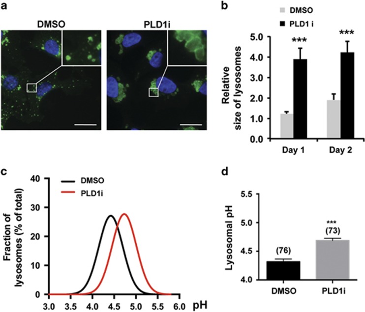
PLD1 inhibition impedes lysosomal functions. (a) PLD1 inhibition caused prominent lysosome enlargement. MDA-MB-231 cells were grown in low glucose medium and treated with DMSO or PLD1i for 2 days. Cells were fixed and then permeabilized with saponin that preserved good lysosomal morphology before staining for LAMP1 (green) and DAPI for nuclei (blue) (scale bar: 20 μm). (b) Quantification of lysosomal size in MDA-MB-231 cells as in (a). Lysosome size was determined by the areas of LAMP1-labeled vesicles using ImageJ; n=3. (c) PLD1 inhibition increased lysosomal pH. Distribution histograms of lysosomal pH values (fitted to Gaussian distributions) measured by ratiometric imaging from DMSO and PLD1i-treated MDA-MB-231 cells grown for 2 days in low glucose medium. (d) Mean lysosomal pH values of data points in (c); n=3. ***P<0.001

