Figure 5.
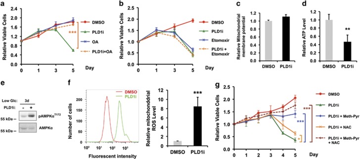
PLD1 regulates free FA generation to support mitochondrial FAO in MDA-MB-231 cells exposed to low glucose medium. (a) Addition of exogenous oleic acids fully rescued cell death induced by PLD1i; n=3. (b) Etomoxir, a FAO inhibitor, reduced viability of MDA-MB-231 cells similarly as PLD1i. The combined treatment with etomoxir and PLD1i had no additive effect; n=3. (c) PLD1i had no effect on mitochondrial membrane potential; n=3. (d) PLD1i reduced ATP production; n=3. (e) PLD1i treatment increased the phosphorylation of AMPKα on day 3 in low glucose medium. (f) PLD1i increased mitochondrial ROS level. The MDA-MB-231 cells stained with MitoSOX Red mitochondrial ROS indicator were analyzed by flow cytometry. Left, histograms of MitoSOX fluorescent intensity. Right, statistic results of the relative mitochondrial ROS level; n=3. (g) The cell death phenotype of PLD1i-treated cells was rescued by methyl-pyruvate (meth-Pyr) and N-acetylcysteine (NAC); n=3. *P<0.05; **P<0.01; ***P<0.001

