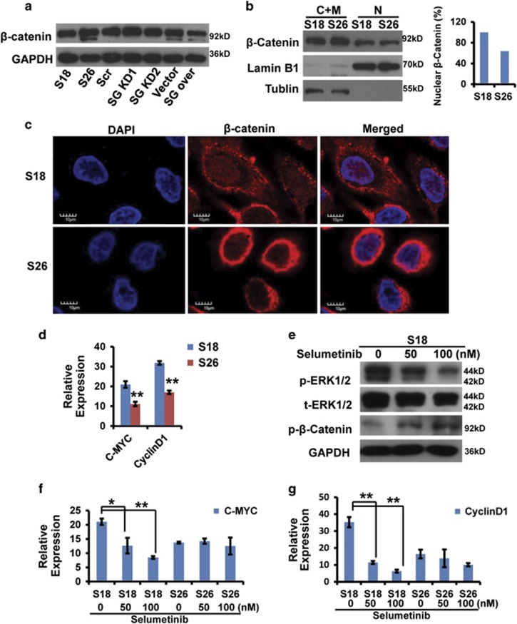
Figure 6.
β-Catenin localization in S18 and S26 cells. (a) β-Catenin protein levels in the indicated cells determined by western blot analysis. (b) Nuclear (N) and cytosolic/membrane (C+M) fractions of β-catenin in S18 and S26 cells determined by western blot analysis. Lamin B1 and tubulin were used as controls for the N and C+M compartment, respectively. (c) Localization of β-catenin in S18 and S26 cells by confocal immunofluorescence analysis (× 400). (d) c-Myc or cyclinD1 mRNA expression (normalized to GAPDH) in S18 and S26 cells. Data represent the average±S.D., n=3; **P<0.01 for S26 cells compared with S18 cells. (e) Western blot analysis of whole-cell lysates from S18 cells. (f and g) c-Myc or cyclinD1 mRNA expression (normalized to GAPDH) in S18 and S26 cells treated with selumetinib (ERK inhibitor) for 48 h. Data represent the average±S.D., n=3; *P<0.05, **P<0.01 versus 0 nM-treated cells

