Figure 7.
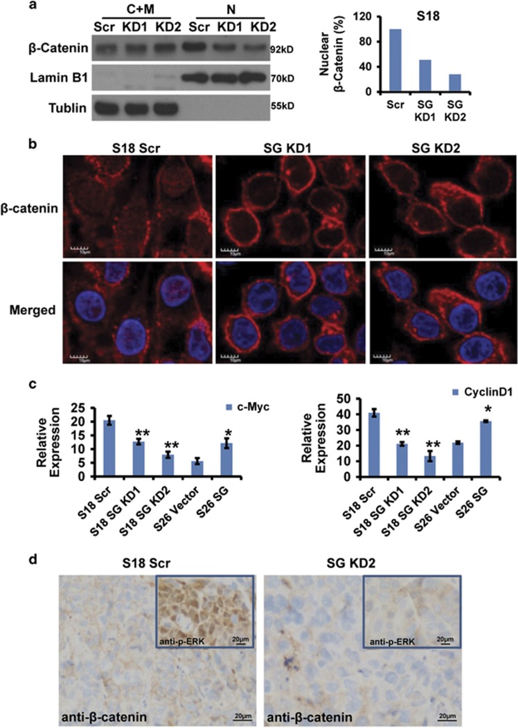
Serglycin induced β-catenin activation. (a) Nuclear (N) and cytosolic/membrane (C+M) fractions of β-catenin in S18 scrambled, S18 SG KD1 and S18 SG KD2 cells determined by western blot analysis. (b) Localization of β-catenin in S18 scrambled, S18 SG KD1 and S18 SG KD2 cells by confocal immunofluorescence analysis (× 400). (c) c-Myc mRNA expression (normalized to GAPDH) in the indicated cell lines. Data represent the average±S.D., n=3; *P<0.05, **P<0.01 versus DMSO-treated cells. (d) IHC staining of β-catenin and p-ERK1/2 in S18 scrambled tumor tissues and S18 SG KD2 tumor tissues (200 ×)

