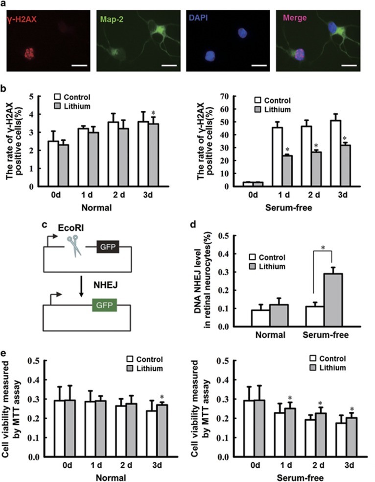
Figure 1.
Lithium pretreatment of cultured ischemic retinal neurocytes decreases DNA damage by increasing NHEJ repair in vitro. (a) Retinal neurocytes were stained with Map-2 (green) and γ-H2AX (red) at different time points. The relative quantification of γ-H2AX expression following serum starvation was determined by counting foci in 50 randomly selected, Map-2-positive cells. Scale bars: 20 μm. (b) lithium exposure caused a marked decrease in γ-H2AX staining (0 day, 3.03±0.27% 1 day, 23.64±1.11% 2 days, 26.47±1.67% 3 days, 31.78±2.15%) compared with the control under a serum-free condition from the first day (0 day, 3.00±0.19% 1 day, 45.51±4.44% 2 days, 46.50±4.76% 3 days, 50.99±5.17% *P<0.05). Data are graphically represented with normal and serum-free condition. n=3 for each group, *P<0.05. (c) Schematic of the NHEJ repair assay. (d) Rejoining capacity of retinal neurocytes with or without lithium. The plasmid substrate pEGFP-N1 was linearized with the EcoRI endonuclease, which cleaves between the promoter and the GFP gene and prevents GFP expression. Recircularization of the linearized plasmid was detected as GFP-positive signal via flow cytometric analysis. n=3 for each group, *P<0.05. (e) The MTT assay showed that lithium treatment also increased viability on the third day (3d) (3 days: control, 0.238±0.054; lithium, 0.269±0.015). However, in neurocytes deprived of serum, lithium-treated improved cell survival from the first day (1 day, 0.251±0.032; 2 days, 0.225±0.031; 3 days, 0.202±0.026) compared with the serum-starved controls that were not treated with lithium (1 day, 0.228±0.049; 2 days, 0.192±0.025; 3 days, 0.175±0.041). n=3 for each group, *P<0.05. All error bars represent SEM

