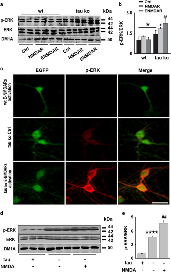
Figure 4.
Tau deletion restores ERK activation in E-NMDAR-activated primary mouse cortical neurons and hippocampus. (a) Wt- or tau-deleted mouse neurons were cultured and treated with synaptic or extrasynaptic NMDAR activating protocols for 24 h, respectively. The total protein levels and active forms of ERK were detected by western blotting. (b) Quantitative analysis of the protein levels in (a), *P<0.05 versus wild-type neurons, #P<0.05 versus tau Ko control neurons, ##P<0.01 versus tau Ko control neurons, n=6, N=3 independent cultures. (c) Wt or tau Ko mouse neurons were cultured and treated with or without E-NMDAR-activating protocols for 24 h, p-ERK and EGFP staining were acquired by confocal microscopy, for Wt neurons, the cells were transfected with EGFP lentivirus to be visualized. Scale bar=50 μm. (d) Wt or tau Ko mice were injected with saline (NS) or NMDA (60 mM, 2 μl) into the hippocampus. Twenty-four hours later, left part of hippocampi was isolated and homogenized, total protein levels and active forms of ERK were detected by western blotting. (e) Quantitative analysis of the ERK levels in (d), ****P<0.0001 versus Wt neurons, ##P<0.01 versus tau Ko control neurons, n=3 mice per group

