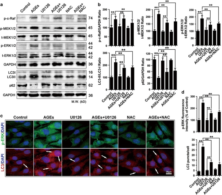
Figure 5.
AGE-induced mesangial cell autophagy was mediated by the ROS/ERK signaling pathway. (a–d) Cells were pre-treated with or without U0126 (30 μM) or NAC (5 mM) and incubated with AGEs (250 mg/l) for 24 h. (a) Western blot analysis of p-c-Raf, p-MEK1/2, t-MEK1/2, p-ERK1/2 and t-ERK1/2 protein expression levels and autophagy-related protein (LC3II/I and p62) levels in mesangial cells. (b) Quantitative analysis of p-c-Raf, p-MEK1/2, p-ERK1/2, LC3II/I and p62 protein expression levels. The data are presented as the mean±S.E.M. from at least three independent experiments. **P<0.01. (c) LC3/p-ERK expression was assessed by fluorescent microscopy. Arrowhead: LC3 puncta accumulation. Bar=2 μm. (d) Quantitative analysis of p-ERK fluorescence intensity and LC3 puncta. The data are presented as the mean±S.E.M. from at least three independent experiments. **P<0.01

