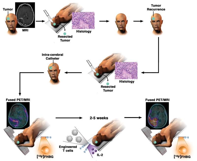
Fig. 2. Schematic demonstrating the workup and monitoring of CTL infusion after an initial treatment for high-grade glioma.
After enrollment in this study and upon tumor recurrence, an intracerebral Rickham catheter was installed for repetitive infusions of CTLs. Each patient underwent [18F]FHBG PET and MRI before and after CTL infusions. In addition, IL-2 was administered at regular intervals to further increase the survival and enhance the potency of the administered CTLs. CTL distribution was imaged by measuring changes in tumoral [18F]FHBG accumulation before and after infusions of cells.

