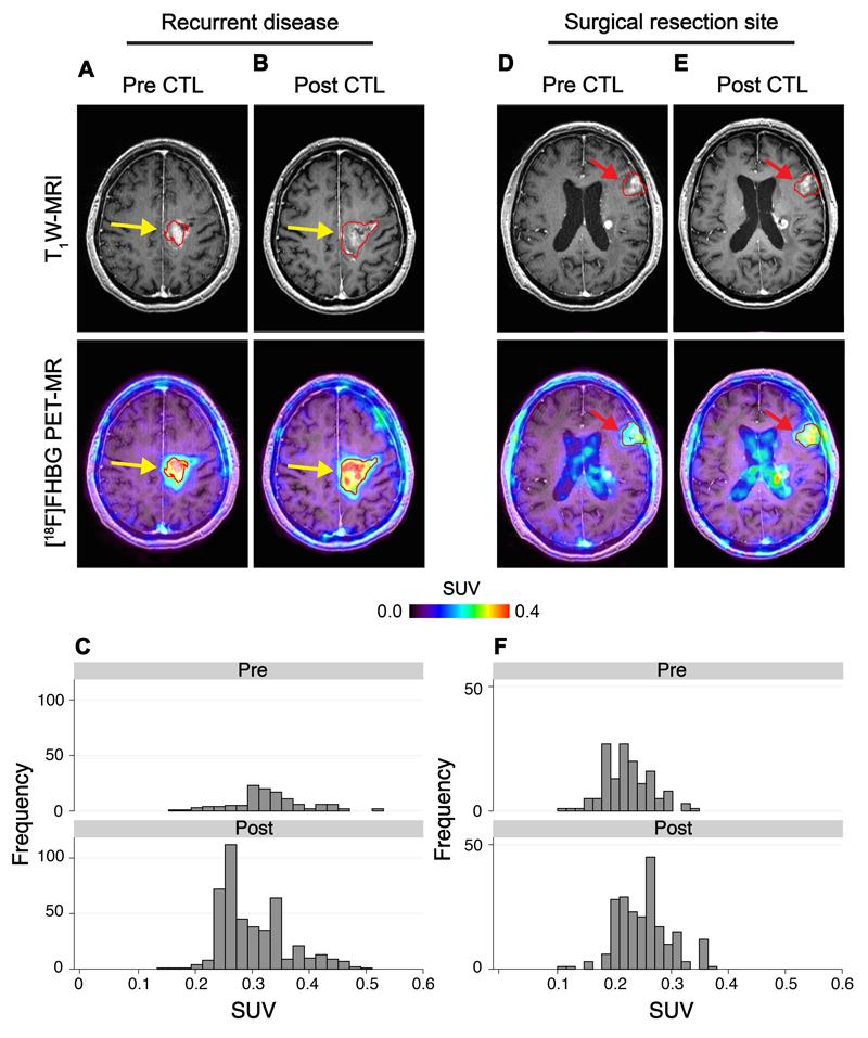
Fig. 4. [18F]FHBG PET imaging in recurrent disease and at untreated tumor sites.
[18F]FHBG PET imaging was performed in a 60 year-old male (Patient 7) with multifocal left hemispheric glioma. CTLs were injected into the medial left frontal lobe tumor (yellow arrows). (A) Tumor size was monitored by T1-weighted (T1W) contrast-enhanced MRI (top left panels). [18F]FHBG PET images were fused with MRI images (bottom left panels), and 3D volumes of interest were drawn using a 50% [18F]FHBG SUVmax threshold, outlined in red. (B) MRI and [18F]FHBG PET-MR images one week after CTL infusions. (C) Voxel-wise analysis of [18F]FHBG total radioactivity in pre- and post-CTL infusion scans. [18F]FHBG activity was additionally assessed in a non-injected tumor focus (red arrows) before (D) and after (E) CTL infusions, with voxel-wise analysis of this lesion performed for comparison (F).

