Figure 2.
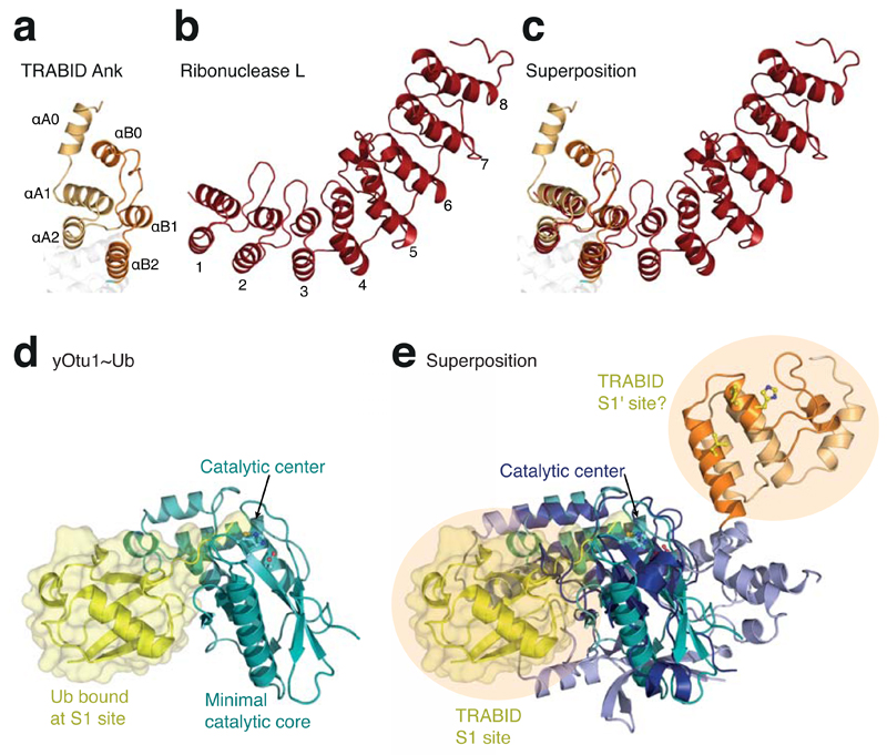
TRABID comprises two Ankyrin repeats with roles in Ub binding. (a) Structure of the Ank domain in TRABID showing the two repeats. (b) Structure of ribonuclease L (pdb-id 1wdy, 47), the Ank repeat protein with highest similarity to the TRABID Ank domain in a DALI search (Z-score 8.4). The eight Ank repeats are numbered. (c) Superposition of the TRABID Ank domain and ribonuclease L. (d) The minimal OTU domain of yOtu1 (green) with Ub (yellow) bound at the S1 site (pdb-id 3by4, 25). The orientation matches that of the minimal OTU domain core indicated in Fig. 1c. (e) Superposition of TRABID and yOtu1 reveals the relative position of the S1 Ub binding site on TRABID, and suggests that the Ank domain may constitute an S1' Ub binding site.

