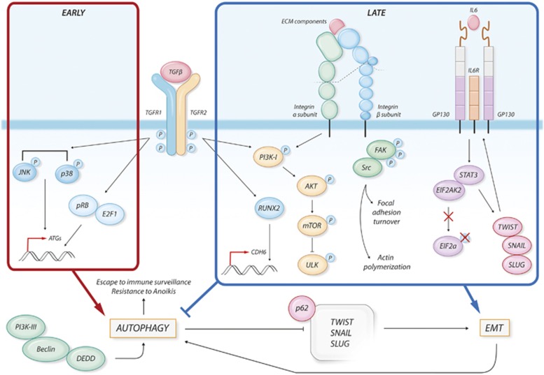
Figure 4.
Signaling pathways at the crossroad between autophagy and EMT. During cancer progression, TGFβ activates several pathways. In the early phases of tumor development, TGFβ promotes the expression of pro-autophagic genes (ATGs) through the activation of the p38 and JNK pathways and through the induction of pRB/E2F1 transcriptional activity. On the other hand, during later phases of tumor progression, TGFβ-mediated activation of the PI3K–AKT–mTOR pathway has been linked to promotion of EMT and to inhibition of autophagy through inhibition of ULK1. During EMT, TGFβ also induces the expression of the mesenchymal marker CDH6, through the activity of the RUNX2 transcription factor. Changes in the ECM interaction properties of the cells lead to Integrin activation. the PI3K–AKT–mTOR and FAK–SRC axis are activated by Integrins and mediate the translation of mechanical and environmental stimuli within the cells. The FAK–Src pathway leads to FA turnover and actin polymerization, promoting the acquisition of a mesenchymal phenotype. IL-6 overexpression results in the activation of STAT3 that is able to sequestrate EIF2AK2, not allowing the phosphorylation of the pro-autophagic factor eIF2α. Moreover, STAT3 induces the expression of the EMT-promoting transcription factors TWIST, SNAIL and SLUG, also stimulating an autocrine signal through the upregulation of IL-6. Autophagy and EMT can also regulate each other. DEDD interacts with the autophagic-promoting complex PI3K-III/BCN1, leading to autophagy-mediated degradation of TWIST, SNAIL and SLUG thus attenuating EMT. In turn, EMT can enhance autophagy to help survive stressful conditions, escape immune surveillance and overcome cell death

