Figure 5.
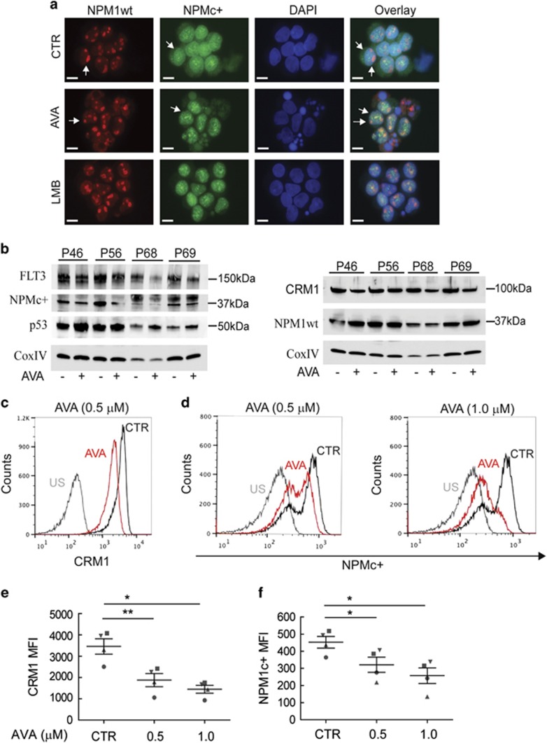
Avrainvillamide treatment decreases expression levels of NPMc+, CRM1 and FLT3 proteins in AML primary patient cells. (a) Four primary AML samples (Supplementary Table 5) were treated with DMSO (CTR), avrainvillamide (AVA; 0.5 μM and 1.0 μM) for 24 h or LMB (10 nM) for 3 h. Cells were then cytospun onto coverslips, fixed and stained using NPMc+- and NPM1wt-specific antibodies. Results from one representative patient sample (P46) are shown (DAPI indicates nuclei; scale bar, 10 μm). (b) AML patient samples (Supplementary Table 4) were treated with DMSO (CTR), AVA (0.5 μM) for 24 h or LMB (10 nM) for 3 h, lysed and immunoblotted for CRM1, NPMc+, p53, FLT3, NPM1wt and CoxIV expression (loading control). (c–f) AML patient cells (Supplementary Table 5) were treated with DMSO (CTR) or AVA (0.5 μM and 1.0 μM) for 24 h, fixed and analyzed by multiplexed flow cytometry for CRM1 (c), and NPMc+ (d) expression by gating on living cells (stained with an aminoreactive dye before permeabilization), specifically analyzing AML blasts, as determined by the use of anti-CD45- (negative) and anti-CD33- (positive) specific antibodies. Results from one representative patient sample are shown. US, unstained. (e) Flow cytometric median fluorescence intensity (MFI) quantification of CRM1 and (f) NPMc+ expression in the four primary AML patient samples, results represent mean±S.E.M. Comparisons between treated and control samples were conducted using a paired Student's t-test (*P<0.05, **P<0.01)

