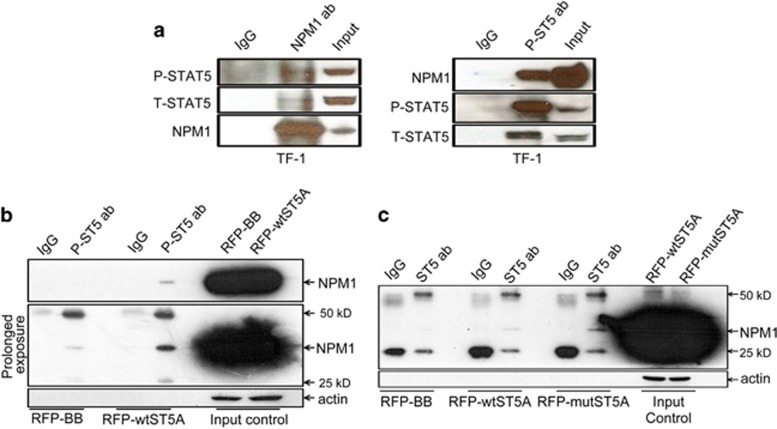
Figure 3.
NPM1 physically associates with STAT5. (a) Lysates from TF-1 cells (4 × 10⁶ cells per condition) maintained in GM-CSF were immunoprecipitated with monoclonal anti-NPM1 antibody (1 μg per condition) and monoclonal anti-phospho STAT5 antibody (1:200) and probed with the indicated antibodies. (b) HEK 293T cells were plated in T-75 culture flasks on day 0 and transfected with either RFP-STAT5A or RFP-backbone vectors at the confluence of 80–90% on day 1, and the transfected cells were harvested and lysed on day 2. Lysate containing 2000 μg total protein was precipitated with 5 μl mouse monoclonal anti-phospho-STAT5 antibody (1:100) or 20 μl mouse IgG sepharose beads, mouse monoclonal anti-NPM1 antibody was used for detection. (c) The transfection of RFP-STAT5AY694F mutant vector followed the same procedure as the preceding panel. The lysate carrying 2000 μg total protein was precipitated with rabbit monoclonal anti-STAT5 antibody (1:50) or rabbit IgG XP Isotype control (1:50), mouse monoclonal anti-NPM1 antibody was used for detection

