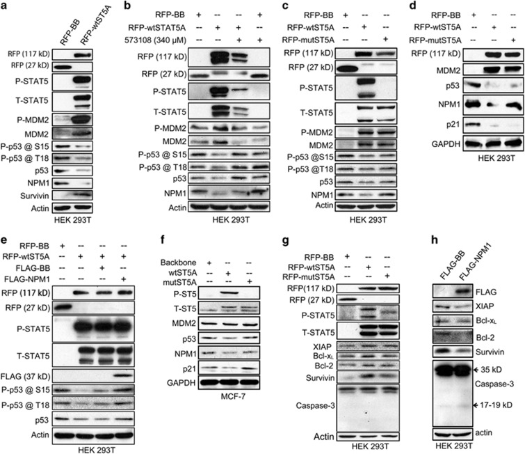
Figure 5.
P-STAT5 regulates p53/MDM2 functions and cell survival through NPM1 protein. (a) HEK 293T cells (6 × 10⁵ per well) transfected with either RFP-STAT5A or RFP-backbone vectors were harvested at 24 h after transfection, and the cell lysates were subjected to immunoblotting assays for detection of expression levels of p53 and its phosphorylation levels at serine 15 and threonine 18, as well as the expression levels of MDM2 and its phosphorylation level at serine 166. (b) Six hours after transfection with either RFP-STAT5A or RFP-backbone vectors, HEK 293T cells (6 × 10⁵ per well) were incubated overnight with inhibitor 573108 at a concentration of 340 μM and then harvested and lysed for immunoblotting assays to examine the expression levels of p53 and MDM2 as well as their relevant phosphorylation levels. (c and d) HEK 293T cells (6 × 10⁵ per well) were transfected with RFP-STAT5AY694F mutant vector for 24 h, harvested and lysed for immunoblotting assays on the expression levels of MDM2, p53 and p21. (e) HEK 293T cells (6 × 10⁵ per well) were co-transfected with RFP-wtSTAT5A and FLAG-NPM1 vectors for 24 h, harvested and lysed for immunoblotting assays on the p53 expression levels. (f) MCF-7 cells (2 × 10⁶ per well) were plated on 10 cm plate on day 0 and then lentivirally transduced with pLVX-IRES-mCherry-wtSTAT5 or pLVX-IRES-mCherry-STAT5Y694F on day 1. The cells were harvested 72 h after the transduction and lysed for immunoblotting assays. (g) HEK 293T cells (6 × 10⁵ per well) were transfected with either RFP-STAT5A or RFP-backbone vectors for 24 h, collected and lysed for immunoblotting assays on the expression levels of XIAP, Bcl-xL, Bcl-2, survivin and caspase-3. (h) HEK 293T cells (6 × 10⁵ per well) transfected with FLAG-NPM1 or FLAG-backbone were subjected to immunoblotting assays on the expression levels of the same proteins indicated in the preceding panel. Immunoblotting data in each panel are representative of at least three independent assays

