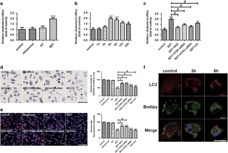
Figure 5.
HY-SDT enhances the lipid catabolism of macrophage via regulating ROS-dependent TFEB nuclear translocation. (a) Cholesterol efflux to HDL (50 μg/well) in macrophage at 6 h after different treatments. (b) Cholesterol efflux to HDL (50 μg/well) in macrophage for the indicated time following HY-SDT. (c) Cholesterol efflux to HDL (50 μg/well) in macrophage pre-treatment with NAC, TFEB siRNA, ATG5 siRNA and CC at 6 h following HY-SDT. (d) Lipids accumulation in macrophage pre-treatment with NAC, TFEB siRNA, ATG5 siRNA and CC was determined by ORO staining. Scale bar=50 μm. (e) Fluorescence microscopy analysis of the uptake of DIL-ox-LDL in macrophage pre-treatment with NAC, TFEB siRNA, ATG5 siRNA and CC at 6 h following HY-SDT. Scale bar=100 μm. (f) Immunofluorescence analysis of lipid catabolism through double fluorescence labeling with Bodipy and anti-LC3 at 3 h and 6 h following HY-SDT. Scale bar=20 μm. *P<0.05, **P<0.01, ***P<0.001 versus control, #P<0.05 versus SDT. All values are given as mean±S.D. (error bars) of three independent experiments

