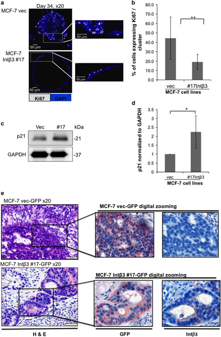
Figure 6.
MCF-7-Intβ3 cells are growth arrested in the 3D BME system and differentiate in vivo. (a–d) MCF-7 cell lines cultured in the 3D BME system. (a) Left panel: representative confocal image of cross section through the middle of an organoid (day 36) stained for Ki67 (white). Right panel: digital zooming of the selected area, white arrow indicates Ki67 positive cells. (b) Quantification of the percentage of Ki67-positive cells within each cross section through the middle of the organoids. Twenty-five organoids of each condition were scored. Bars=50 μm. (c) W.B. analysis for the expression of p21 and its quantification normalized to GAPDH in the organoids (d); (n=3) (e) Serial paraffin section of teratomas injected with either MCF-7-vec-GFP or MCF-7-Intβ3-GFP cells. Paraffin section were subjected to H&E staining (magnification × 20), or subjected to either GFP or Int-β3 staining (digital zooming of the selected area is presented). Representative images; n=3

