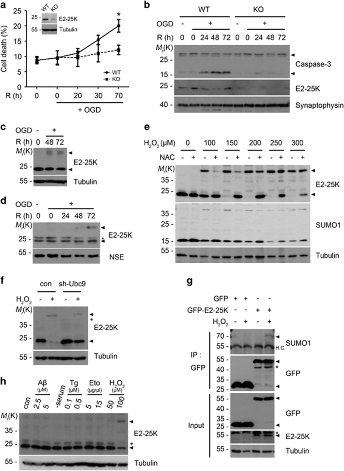
Figure 1.
Neuronal cell death in OGD/R is blocked by E2-25K deficiency and accompanied by E2-25K SUMOylation. (a and b) E2-25K WT or KO mouse primary cortical neurons were cultured for 9 days in vitro (DIV-9) and exposed to OGD for 3 h followed by reoxygenation for the indicated times. Cell death rates were measured by trypan blue exclusion assay and expression level of E2-25K was examined by western blotting (insert). Values represent mean±S.E.M. (n=3, two-way ANOVA followed by Bonferroni's post hoc test, *P<0.05) (a). Cell extracts were examined with western blot analysis (b). (c and d) SH-SY5Y cells (c) and WT mouse primary cortical neurons (DIV-10) (d) were exposed to OGD for 3 h and reoxygenation for the indicated times. Cell extracts were then analyzed by western blotting. (e) SH-SY5Y cells were incubated with the indicated concentrations of H2O2 for 12 h in the presence or absence of preincubation with 500 μM NAC for 1 h and analyzed by western blotting. (f) SH-SY5Y cells were transfected with Ubc9 shRNA for 24 h and treated with 150 μM H2O2 for 22 h. (g) HEK293T cells were transfected with GFP or GFP-E2-25K for 24 h and then exposed to 150 μM H2O2 for 18 h. Cell extracts were prepared and subjected to immunoprecipitation (IP) assay using anti-GFP antibody. The immunoprecipitates and cell extracts (input) were analyzed by western blotting. (h) SH-SY5Y cells were left untreated (con) or treated with Aβ1-42, thapsigargin (Tg), etoposide (Eto) or H2O2 for 16 h or serum-free medium (-serum) for 20 h

