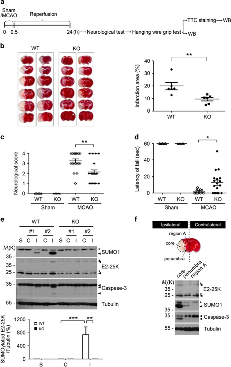
Figure 3.
E2-25K deficiency ameliorates MCAO/R injury. (a) Schematic diagram showing the experimental schedule. (b) The 3–4-month-old male E2-25K WT and KO mice were subject to MCAO for 30 min and reperfusion for 24 h. The 2-mm coronal brain sections, which were prepared from the olfactory bulb to the cerebellum, were analyzed after TTC staining (n=2 for each genotype in this figure; left). Bars represent the percentage of infarction area to whole area with mean±S.E.M., WT mice, n=6; KO mice, n=8 for the analysis, unpaired two-tailed Student's t-test, **P<0.01 (right). (c) The neurological deficits in each mouse were assigned as a score (mean±S.E.M., each sham, n=5; WT mice, n=19; KO mice, n=22, two-way ANOVA followed by Bonferroni's post hoc test, **P<0.01). (d) The mice were analyzed with wire hanging test (mean±S.E.M., each sham, n=5; WT mice, n=10; KO mice, n=18, two-way ANOVA followed by Bonferroni's post hoc test, *P<0.05). (e) Brain extracts (except cerebellum) were prepared from non-ischemic (contralateral, C), ischemic (ipsilateral, I) and sham control (S) hemispheres of E2-25K WT and KO mice and analyzed by western blotting (upper). Bars indicate mean±S.E.M. (lower; n=4, two-way ANOVA followed by Bonferroni's post hoc test, **P<0.01, ***P<0.001). (f) TTC-stained section showing examples of lesions from MCAO/R-treated WT mouse. Brain extracts were prepared from ischemic core, penumbra and the rest of the region (region A) in the ipsilateral region (upper) and analyzed by western blotting (lower). Asterisks indicate non-specific signals

