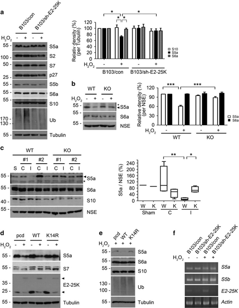
Figure 5.
S5a within proteasome is decreased by SUMOylated E2-25K. (a and b) B103/con and B103/sh-E2-25K cells (a) and E2-25K WT and KO mouse cortical neurons (DIV-20) (b) were treated with 100–150 μM H2O2 for 14–16 h and analyzed by western blotting (a and b; left). The signals of S5a on the blots were quantified by densitometric analysis and normalized by tubulin (a, right) or NSE (b, right). Bars represent mean±S.E.M., n=4, two-way ANOVA followed by Bonferroni's post hoc test, *P<0.05, ***P<0.001). (c) The 3–4-month-old male E2-25K WT and KO mice were perfused for 24 h after MCAO for 30 min. Tissue extracts (except cerebellum) from contralateral (C), ipsilateral (I) and sham control (S) hemispheres were analyzed by western blotting (left). The signals of S5a on the blots were quantified by densitometric analysis and normalized by NSE (mean±S.E.M., n=3, two-way ANOVA followed by Bonferroni's post hoc test, *P<0.05, **P<0.01). (d and e) SH-SY5Y (d) and B103/sh-E2-25K (e) cells were transfected with pcDNA (pcd), E2-25K WT or K14R and treated with 200 (d) or 70 μM (e) H2O2 for 20 h. (f) B103/con and B103/sh-E2-25K cells were treated with 200 μM H2O2 for 14 h. Total mRNA was isolated and subjected to RT-PCR analysis

