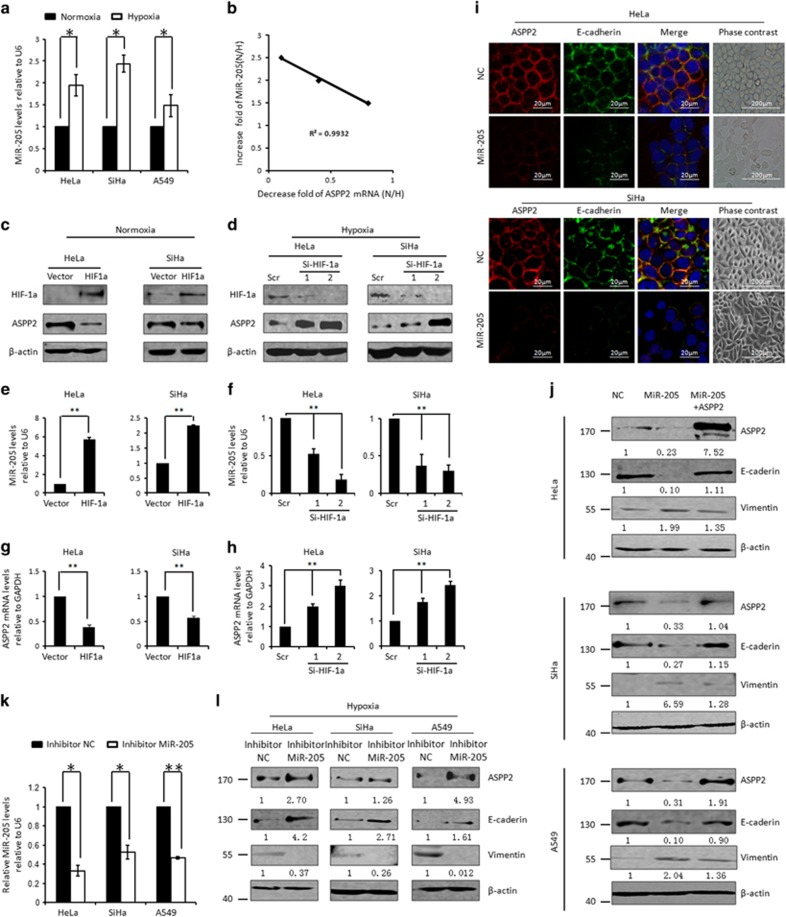
Figure 3.
Hypoxia inhibits ASPP2 by the promoted MiR-205. (a) Real-time RT-PCR assay of MiR-205 under normoxia and hypoxia conditions. Histograms represent the mean±S.E.M. from three independent assays. (b) Linear correlation between decreased ASPP2 and increased MiR-205 was obtained in cancer cell lines (r2=0.9932, P<0.01). (c) WB analysis of HIF-1a and ASPP2 after ectopic expression of HIF-1a under normoxia conditions. β-Actin was used as a loading control. (e and g) Real-time RT-PCR analysis of MiR-205 (e) and ASPP2 (g) under the conditions as described in c. (d) WB analysis of HIF-1a and ASPP2 after preventing hypoxia-induced HIF-1a by transfection with two independent RNAi specifically targeting HIF-1a (Si-HIF-1a 1 and 2). β-Actin was used as a loading control. (f and h) Real-time RT-PCR analysis of MiR-205 (f) and ASPP2 (h) under the conditions as described in d. (i) Immunostaining analysis of E-cadherin/ASPP2 expression and localization after transfection with NC mimic and MIR-205 mimics. (j) WB analysis of ASPP2, E-cadherin and Vimentin after transfection with negative control, MiR-205 mimics or MiR-205 mimics+ASPP2 in HeLa, SiHa and A549 cells. (k) Real-time RT-PCR analysis of MiR-205 after transfection with NC inhibitors or MiR-205 inhibitors under hypoxia conditions. (l) WB analysis of ASPP2, E-cadherin and Vimentin under the conditions as described in k. β-Actin was used as a loading control. All histograms represent the mean±S.E.M. from three independent assays. *P<0.05; **P<0.01

