Figure 5.
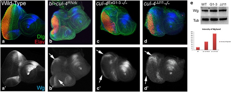
Wg is ectopically induced in cul-4 mutant background. Expression of Wg (blue) in (a,a') Wild-type, (b and b') bi>cul4RNAi, (cul-4 RNAi is misexpressed on DV margin using bi-Gal4), (c and c') cul-4ExG1−3and (d and d') cul-4JJ11 loss-of-function clones. Note robust ectopic Wg (blue) expression on (b') DV margin (marked by white arrows) along with suppression of eye fate. (c and d) The reduced eye phenotype of cul-4 loss-of-function clones generated by cell-lethal approach is accompanied by ectopic upregulation of Wg (blue, marked by white arrow). (a'–d') Shown is the split channel of Wg expression. (e) In western blot analysis, the Wg protein levels are more than twofolds in eye discs with cul-4 loss-of-function clones as compared with wild-type eye disc. Wg band staining intensity calculated by Image-J

