Figure 1.
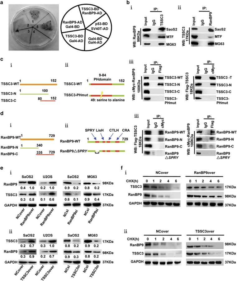
RanBP9 interacts with TSSC3 via post-translational mechanism. (a) Yeast strain Y190 strain was co-transformed with the indicated binding domain (BD) plasmids and activation domain (AD) plasmids. Co-expression of BD-TSSC3 and AD-RanBP9 induced formation of blue colonies on SD/-Trp/-Leu, similarly to positive control cells expressing murine p53 and SV-40 large T-antigen. Gal4-BD and Gal4-AD were used as negative controls. (b) Confirmation of the interaction between endogenous RanBP9 and TSSC3 in osteosarcoma cells. Co-immunoprecipitation assays of whole-cell lysates using anti-TSSC3, or nonspecific IgG and probed with anti-RanBP9 (i), or anti-RanBP9 and probed with anti-TSSC3 (ii). Input samples indicate 10% of pre-immunoprecipitated samples. (c) RanBP9 interacts with TSSC3 PH domain. (i) Schematic illustration of the TSSC3 N- and C-terminal constructs and (ii) PH domain-mutant construct (TSSC3-PHmut, the 49th amino acids serine was mutated to alanine). (iii) Immunoprecipitation assays of 293 T cells co-transfected with the indicated constructs using either anti-Flag or anti-cMyc, followed by immunoblot with anti-cMyc or anti-Flag, respectively. (d) TSSC3 interacts with RanBP9 SPRY domain. (i) Schematic illustration of the RanBP9 N- and C-terminal constructs, and (ii) SPRY domain-deleted construct of RanBP9 (RanBP9△SPRY, △aa 212–333). (iii) Immunoprecipitation assays of 293 T cells co-transfected with the indicated constructs using either anti-Flag or anti-cMyc, followed by immunoblot with anti-cMyc or anti-Flag, respectively. (e) Western blot analysis of TSSC3 and RanBP9 in the indicated RanBP9-overexpressing and RanBP9-knockdown (i) or TSSC3-overexpressing and TSSC3-knockdown (ii) cells. Western blot values were normalized to GAPDH. (f) (i) RanBP9 increases the half-life of TSSC3. SaOS2 cells expressing empty vector (NC) or RanBP9 were treated with cycloheximide (CHX, 100 μg/ml) for the indicated times and subjected to immunoblotting as indicated. (ii) TSSC3 increases the half-life of RanBP9. SaOS2 cells expressing empty vector or TSSC3 were processed as described in Figures 1f (i)

