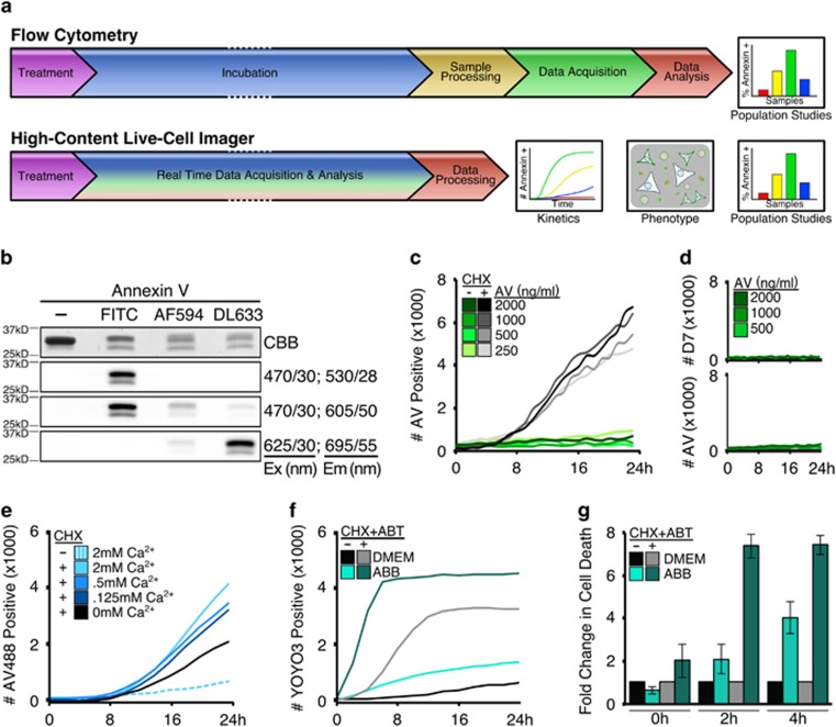
Figure 1.
High-content live-cell imagers provide kinetic real-time Annexin V-binding data without the inherent cell toxicity compared to standard protocols. (a) Annexin V-binding assay workflow by either flow cytometry or high-content live-cell imaging. (b) Recombinant Annexin V analysed by SDS-PAGE and visualized by Coomassie brilliant blue (CBB) or indicated fluorescent filter sets; excitation and emission labelled as filter/bandpass in nm. (c) MEFs were plated, treated as indicated (CHX, 25 μg/ml), incubated with Annexin V-488 (indicated) in growth media, and scanned every hour for 24 h with four frames per well. Events per frame per time point were averaged. (d) MEFs prepared as in (c) and co-incubated with DRAQ7 (600 nM). (e) MEFs were treated/supplemented as indicated (CHX, 50 μg/ml) in the presence of Annexin V-488 (1 μg/ml), and scanned every 2 h for 24 h with one frame per well. Data averaged and representative of at least three experiments. (f) MEFs plated and scanned as in (e), incubated in YOYO3-containing (200 nM) DMEM or ABB, and treated with CHX (50 μg/ml) and ABT-737 (1 μM) to sensitize to apoptosis. (g) Data from (f) converted to fold increase of cytotoxicity in ABB-incubated samples compared to DMEM-incubated samples. Error bars denote standard deviation of triplicate samples

