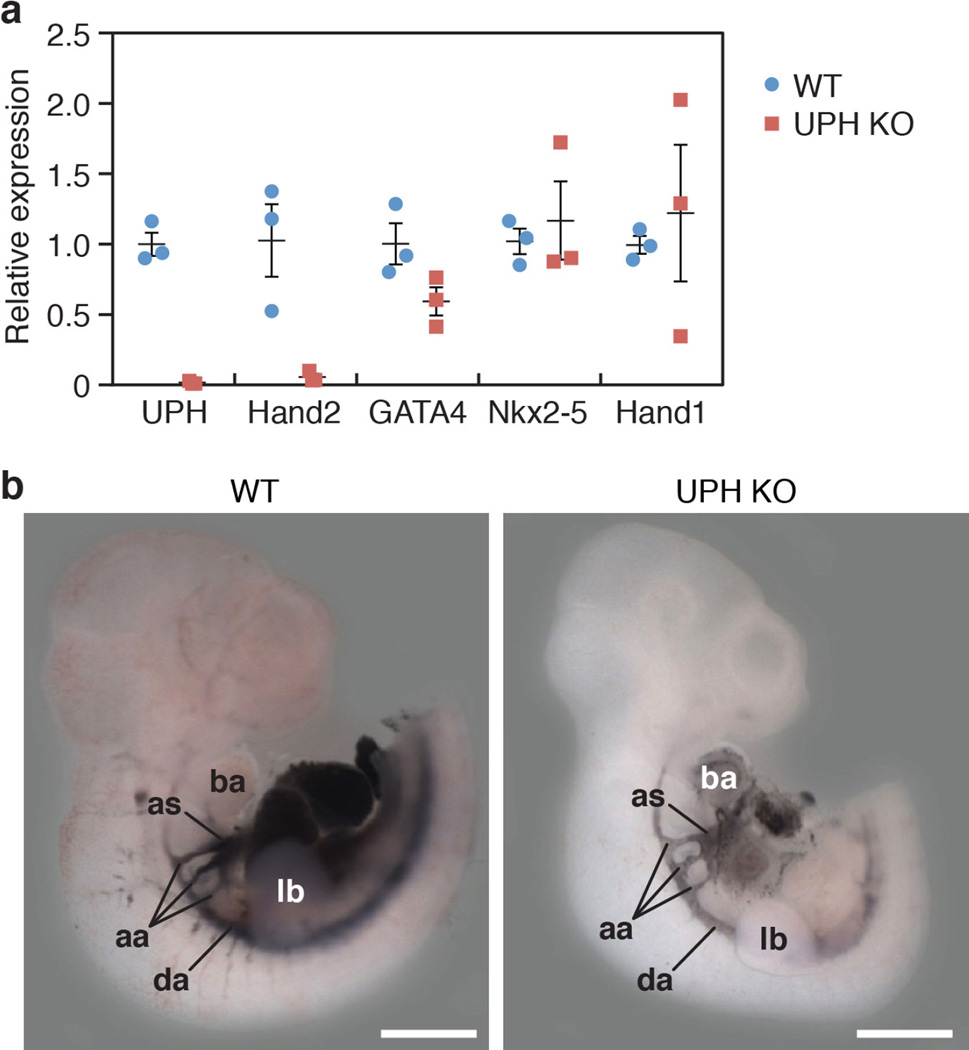
Extended Data Figure 4. Aortic arch arteries are normal in Uph KO embryos.
a, qPCR quantification of gene expression at E10.0 showed robust downregulation of Uph and Hand2 expression in Uph KO hearts, with normal expression of other cardiac transcription factors (n = 3 mice of each genotype from 1 of 3 independent experiments; mean ± s.e.m.). b, India ink was injected into either the left ventricle of wild-type embryos or the single ventricle of Uph KO embryos at E10.5, to visualize the aortic arch arteries and circulation, which appeared normal in Uph KO embryos. aa, aortic arch arteries; as, aortic sac; da, dorsal aorta. Scale bars, 1 mm.

