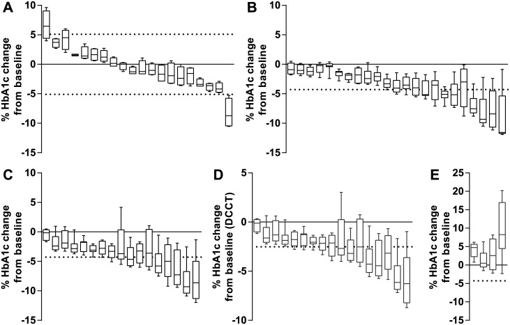
Fig 1. Percentage HbA1c reduction from baseline during 8 weeks post whole blood donation.
Data (Tosoh G8) is shown as box plots each representing one individual. Horizontal dotted lines represent the RCV calculated for SI units. Box plots exceeding the RCV represent a significant change or reduction in HbA1c concentration. A: Control group of 20 non-diabetic volunteers not donating whole blood.9 (Tosoh G8 two-tailed RCV ±5.1%) B: 23 non-diabetic blood donors after 475 mL whole blood donation. (Tosoh G8 one-tailed RCV -4.28%) C: 17 blood donors with type 2 diabetes after 475 mL whole blood donation. (Tosoh G8 one-tailed RCV -4.28%) D: 17 blood donors with type 2 diabetes after 475 mL whole blood donation represented in DCCT units. (Tosoh G8 one-tailed RCV -2.52%) E: 4 blood donors with type 2 diabetes with an increase in HbA1c after 475 mL whole blood donation which were excluded from further analysis. (Tosoh G8 one-tailed RCV -4.28%)

