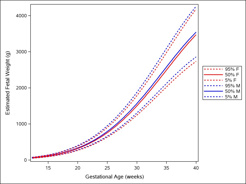
Fig 2. Female and male growth of estimated fetal weight during gestational weeks 14–40.
The difference in growth for female (F; red) and male (M; blue) fetuses is shown by the 5th, 50th, and 95th percentiles for EFW growth. The smoothed lines are based on quantile regression that includes data from all the participating countries.

