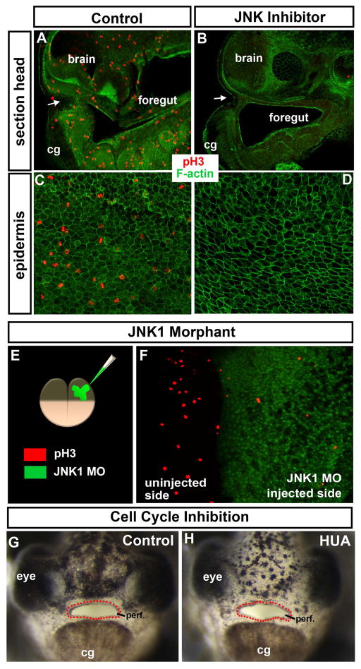
Figure 3.
A–D) Shows phospho-histone H3 (ph3; red) marking mitotic cells and labeling of F-actin with phalloidin (green) is used as a counterstain. White arrows indicate the embryonic mouth. A–B) Sagittal section through the middle of the embryo at stage 37/38 after treatment with DMSO/control (A) or JNK inhibitor (B). C, D) Epidermis at stage 37/38 after treatment overnight with DMSO/control (C) or JNK inhibitor (D). E) Schematic showing the morpholinos injected into one cell at the two cell stage and the key where the morpholino can be seen in green and ph3 is labeled in red. F) Dorsal view of an embryo injected as shown in E. G, H) Frontal views of embryos treated with DMSO (control; G) or cell cycle inhibitors hydroxyurea and aphidicolin (HUA; H). Red dots outline the embryonic mouth. Abbreviations; cg = cement gland, MO = morpholino, ph3= phospho histone H3, perf. =perforated.

